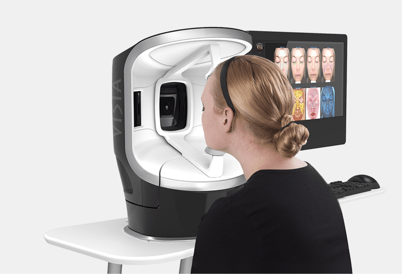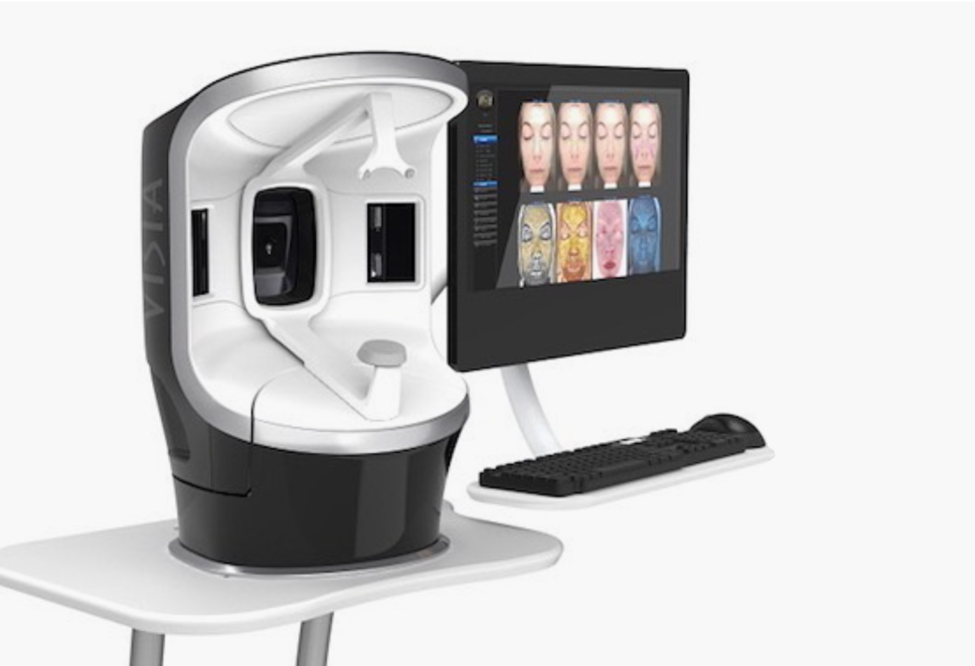


Skin analysis with Visa Gen7
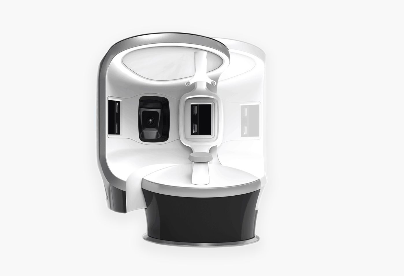
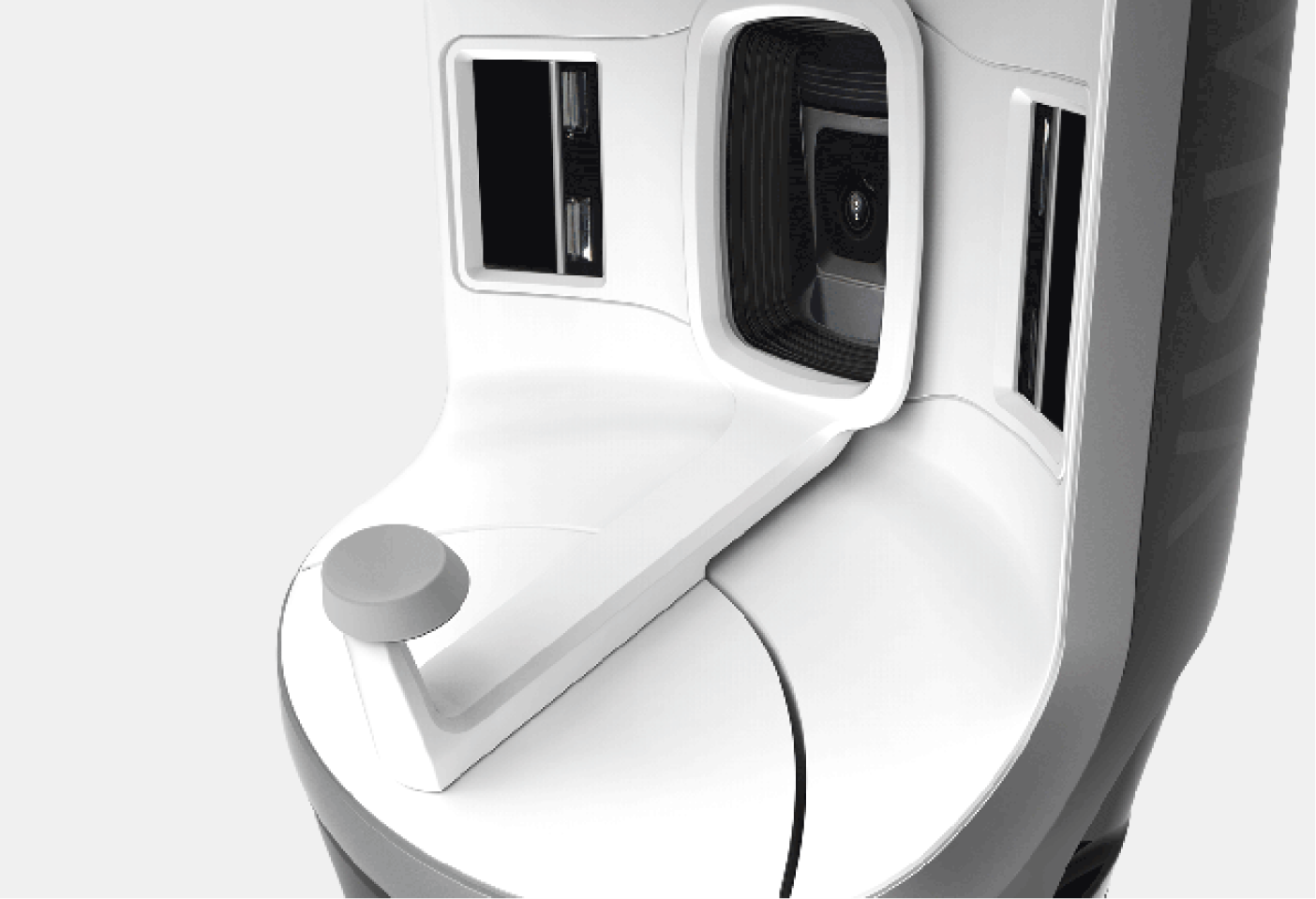
Our advanced skin analysis system VISIA is now also available for the cosmetic-aesthetic area. It was developed by a world leader in 2D and 3D photography systems and is based on extensive scientific research and analysis of treatment results. The system uses Canon's state-of-the-art camera systems and specially developed lighting components to accurately display eight different features of the skin. Here you will find an overview of all features.
- Determine the current condition of your skin
- Determine the relative age of your skin (comparison with average values from a large skin database)
- Show the simulated aging process 5-7 years younger or older (wrinkles and age spots)
- Show you what you will look like in a few years
- Visualize the surface of your skin in 3D
- Make before-and-after comparisons in 3D views
- Simulate injections and volume build-up
- Measure the length and volume of your eyelashes
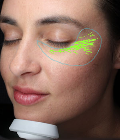
Fold
Wrinkles are skin folds, grooves or creases that occur due to excessive sun exposure and are associated with the skin's decreasing level of elasticity. The extent of wrinkles varies from image to image as it depends on the individual facial expression of the patient/customer. Folds are characterized by their long and narrow shapes.
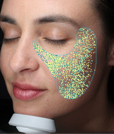
Flatness
Texture analysis primarily assesses the evenness of the skin. The “Evenness” module captures color tone and uniformity by identifying shades of color and detecting the peaks (shown in yellow) and valleys (shown in blue) on the skin surface that indicate variations in surface texture.
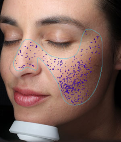
Pores
Pores are round openings of sweat gland ducts on the surface of the skin. Due to shadow effects, pores appear darker than the surrounding skin tone and can be identified by their darker color and round shape using the VISIA system. The VISIA system distinguishes pores from spots based on their size, as pores are much smaller.
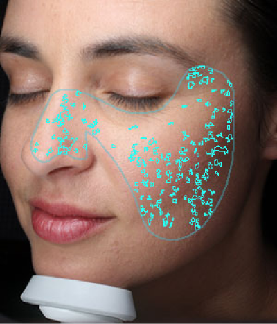
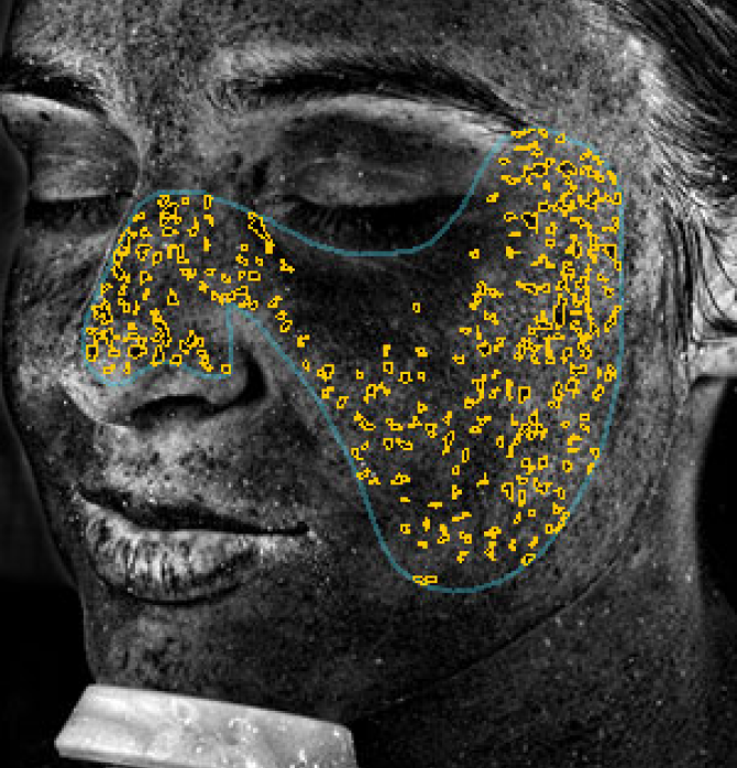
Stains
Spots are brown or red skin lesions that include freckles, acne scars, hyperpigmentation, and vascular lesions. They differ from normal skin tone in their color intensity and contrast. Spots can vary in size and are often circular.
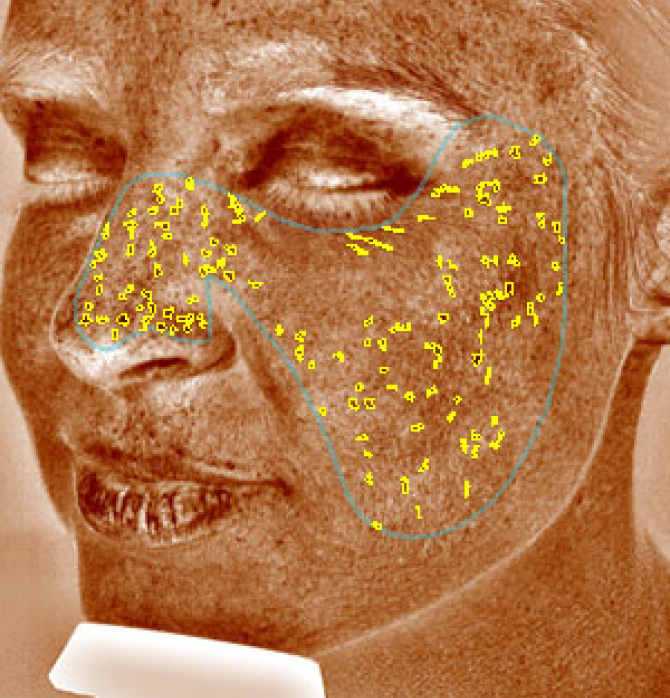
UV stains
UV spots occur where melanin clots beneath the surface of the skin. Under normal lighting conditions, UV spots are usually invisible. However, they are made visible and detected by the VISIA system through the selective absorption of UV light by the melanin in the epidermis.
Brown Spots
Brown spots indicate an increased concentration of melanin on or under the skin. They can represent various forms of hyperpigmentation, such as sun damage or melasma.
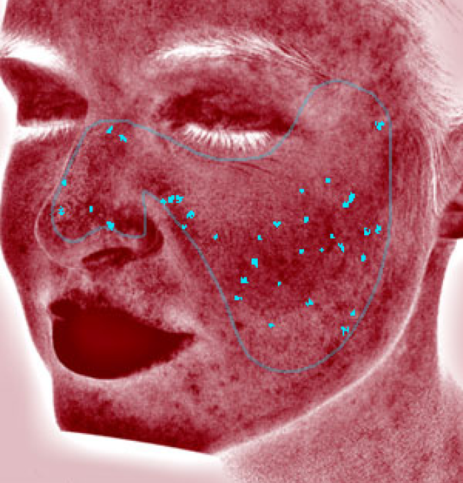
Red Areas
Red areas represent blood or hemoglobin. The evaluation reveals areas beneath the skin's surface that may show signs of vascular diseases such as rosacea or acne.
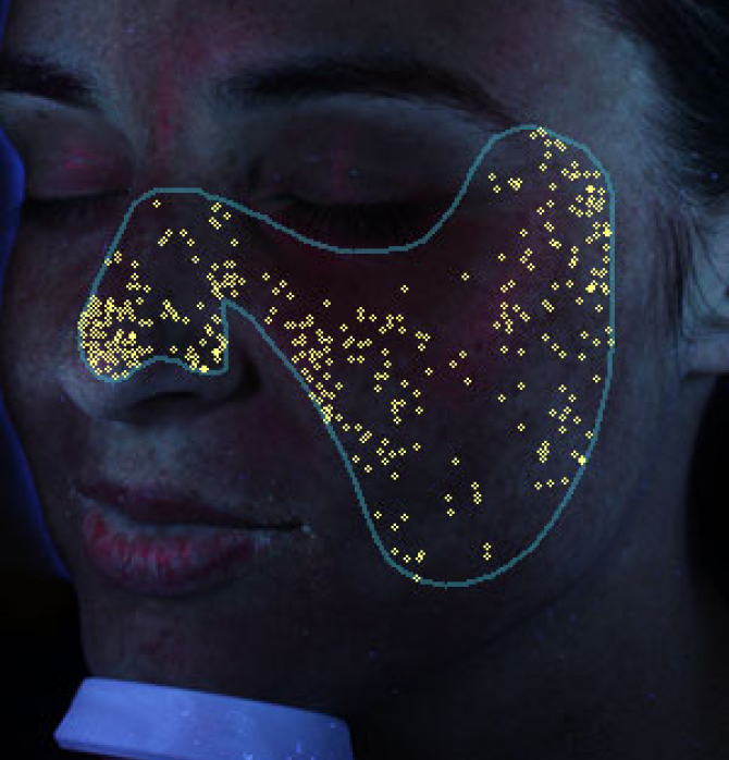
Porphyrine
Propionibacterium acnes, the bacterium that causes acne, produces and stores large amounts of porphyrins that are deposited in the sebaceous glands. These porphyrins fluoresce under UV light and have a circular, shimmering white appearance.
Gallery
Thanks to VISIA Gen7, we can precisely visualize and document the progress of treatments and skin therapies.